|
2 |
|
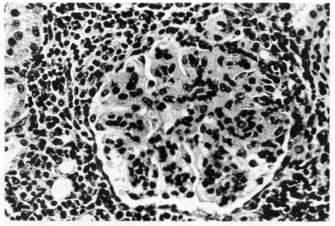
Fig 1—Affected glomerulus Irom a mink with AD. The glomerulus is avascular
with large amounts of eosinophilic granular material. Periglomerular infiltration
of plasma cells and a few lymphocytes are also evident.
quently, subepithelially; they most likely represent deposition of antigen-
antibody complexes.5’6 In some animals viral antigen can also be demon-
strated in the glomeruli. Mononuclear cells proliferate in the liver, begin-
ning in the portal areas but later involving most of the parenchyma. Early
in the disease these cells are a mixed population of lymphocytes, histiocytes,
and immature plasma cells. Later, mature and immature plasma cells domi-
nate. Bile duct proliferation and dilation also occur. Widespread necrotizing
arteritis involving the small muscular arteries are observed in approximately
one-fourth of the spontaneous cases (Figure 3). y-Globulin, CS and viral
antigen are deposited in the affected arteries, suggesting that deposition of
antigen-antibody complexes is the causal factor of the arteritis. All of the
above lesions occur in all mink genotypes when disease is manifest, but their
development is much slower in non-Aleutian type mink. If the latter suc-
cumb to AD, the lesions are just as severe as those in Aleutian mink.
The disease is readily transmitted from mink to mink by parenteral injec-
tion of blood or tissue hemogenates.8 Since the agent is filterable and sedi-
mentable, it appears to be a virus; however, electron micrographs have
failed to reveal valid particles. The agent attains its highest titers 8 to 10
days after inoculation.9,10 A vigorous antibody response against viral antigen
is induced, and it appears that the hypergammaglobulinemia characteristic
of this disease represents antibody.9” Infectious virus complexed with anti-
body circulates and initiates some of the lesions.12 The plasma cell prolifera-
tion is apparently the result of chronic antigenic stimulation and does not
represent a neoplastic process in the classic sense.’3 Immunosuppression of
experimentally infected mink by cyclophosphamide prevents the lesions, but
|
|
2 |